
Before & After Gallery
Mohs Surgery Case #1


About this case
This forehead case shows the progression from before Mohs surgery through reconstruction with a rotation flap. The photographs include the area before treatment, the wound after Mohs surgery, and the repair immediately after reconstruction.
Follow-up photographs at one week and two months document the appearance during healing. View the sequence in order to compare each stage of this patient’s recovery.
Mohs Surgery Case #2


About this case
This nasal case documents Mohs surgery followed by reconstruction with a dorsal nasal rotation flap. The sequence shows the treatment area before surgery, the wound after Mohs surgery, and the completed reconstruction.
Two views taken at the three-month follow-up show the appearance of the repair from different angles. These photographs document this patient’s individual result.
Mohs Surgery Case #3


About this case
This chin case shows Mohs surgery followed by a linear repair. The photographs document the area before treatment, the wound after Mohs surgery, and the closure immediately after reconstruction.
A ten-month follow-up photograph provides a later view of the healed area. Compare the images in sequence to see how the appearance changed over time.
Mohs Surgery Case #4
About this case
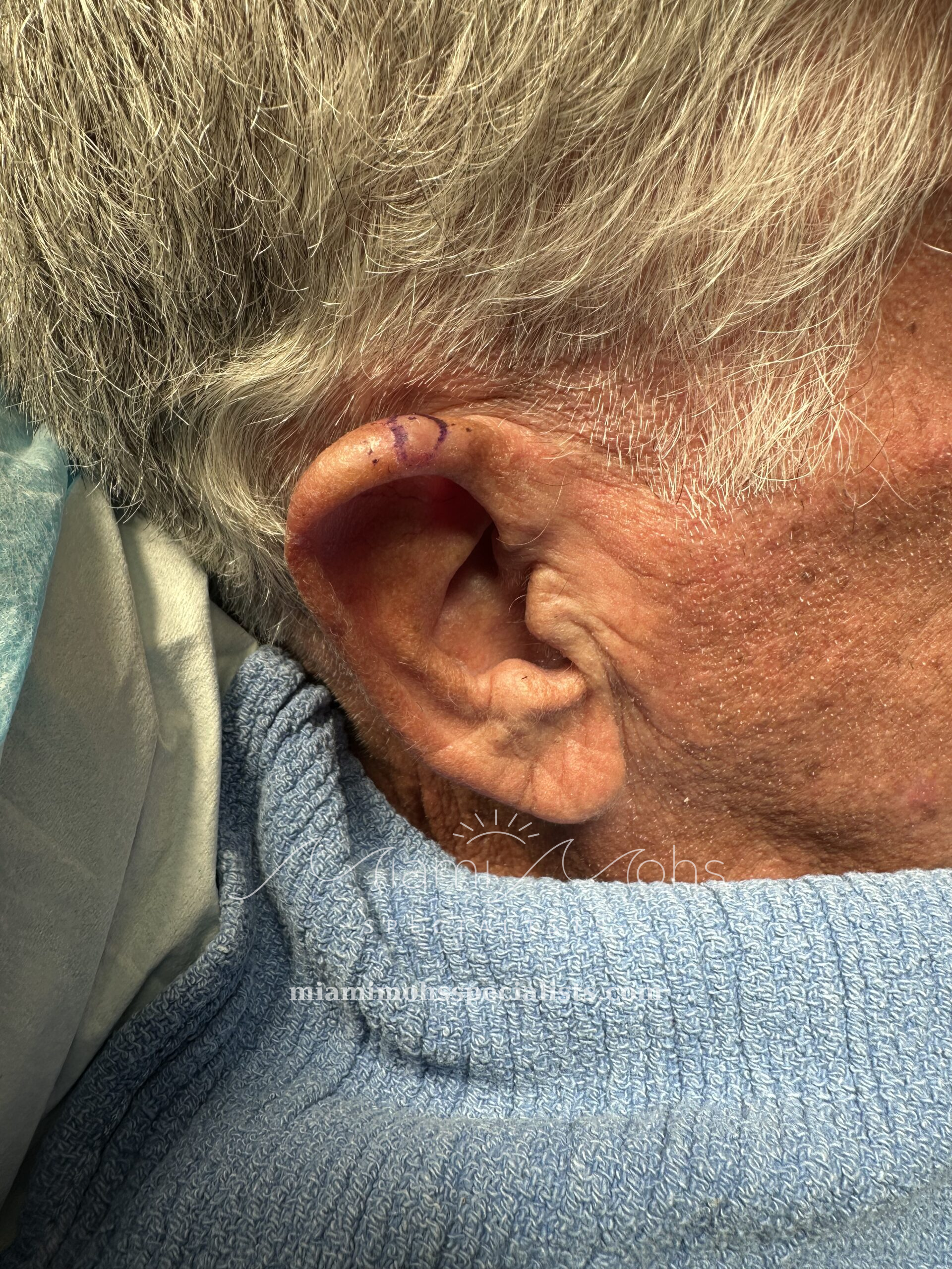
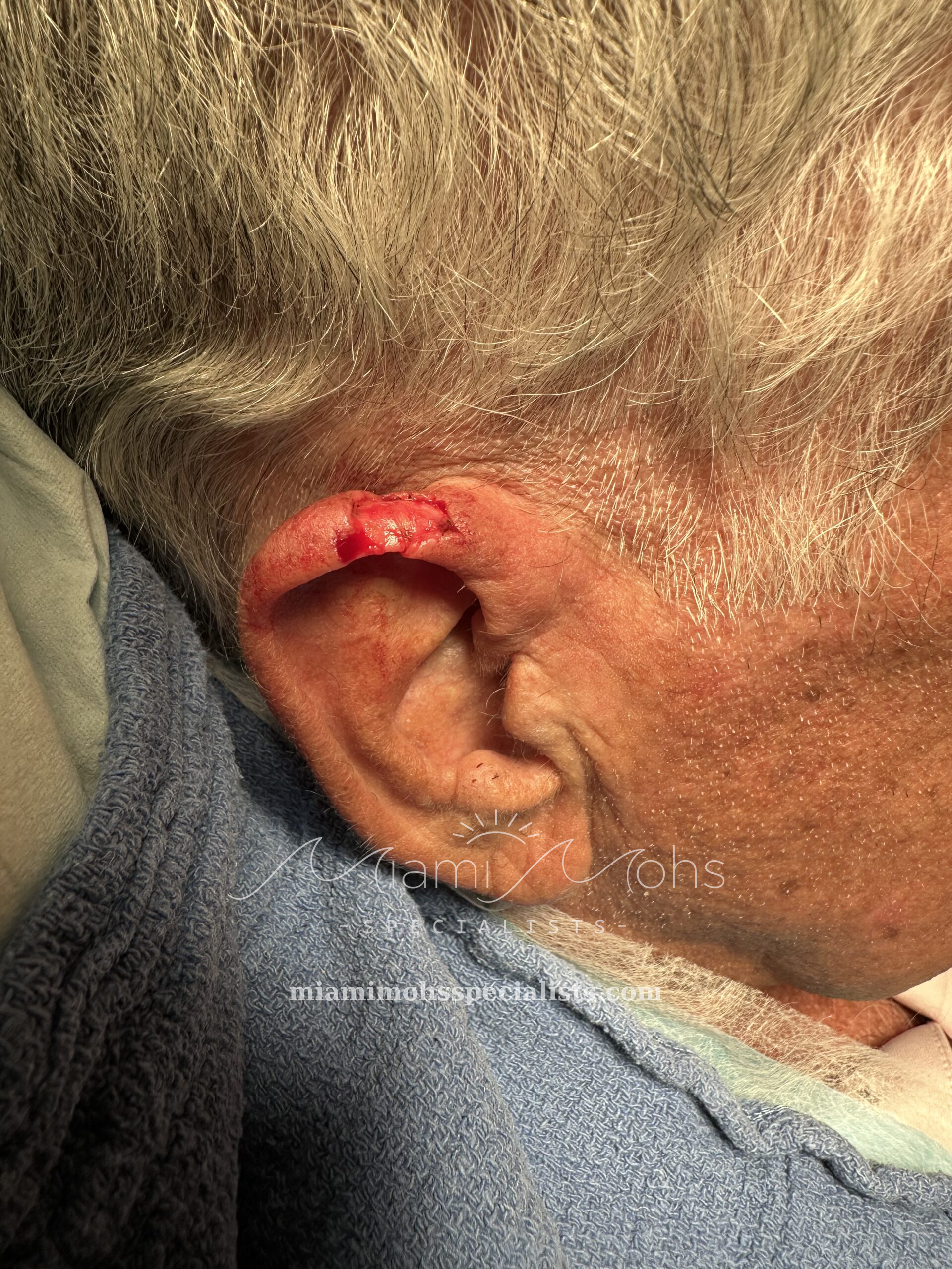
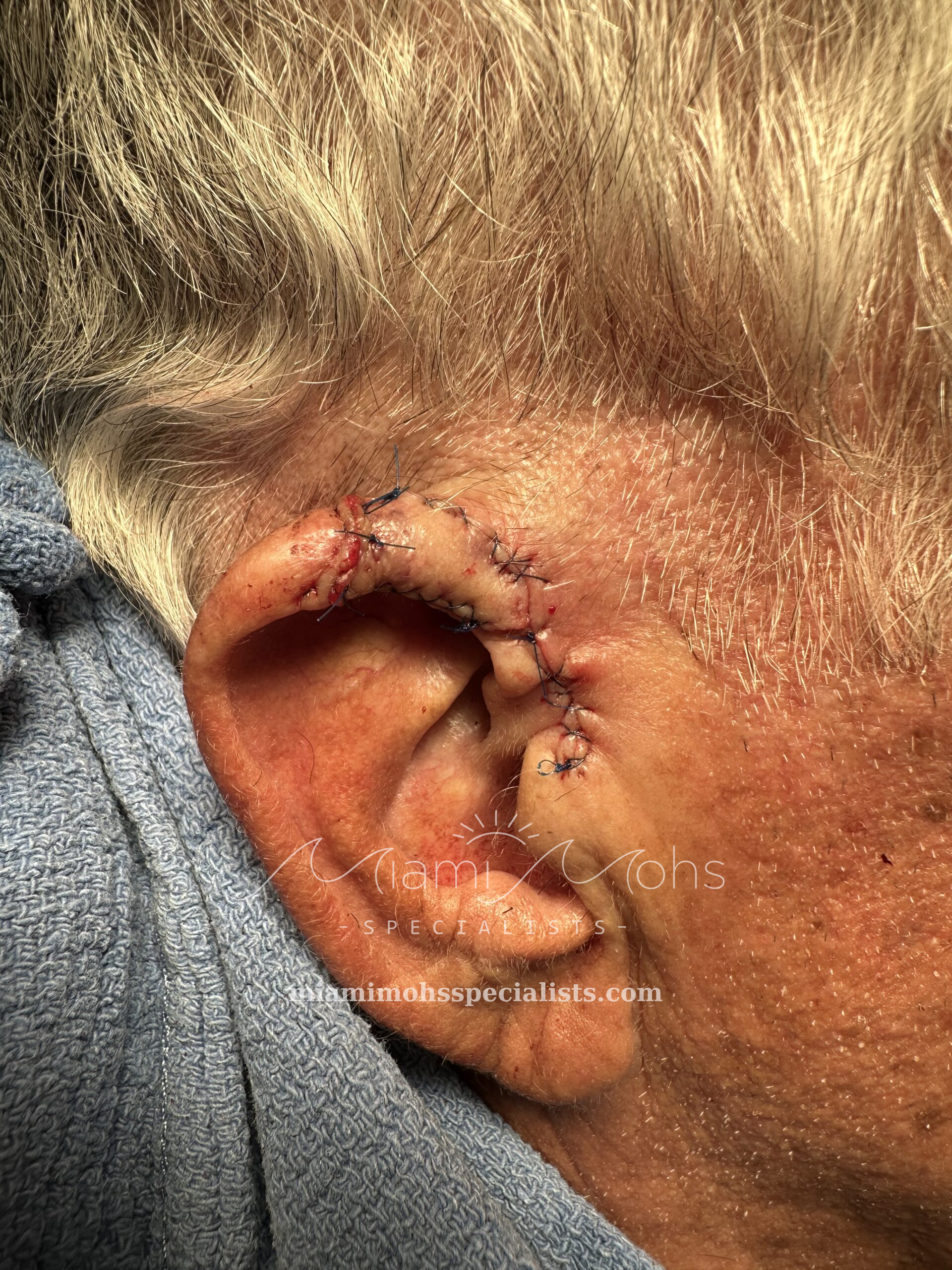
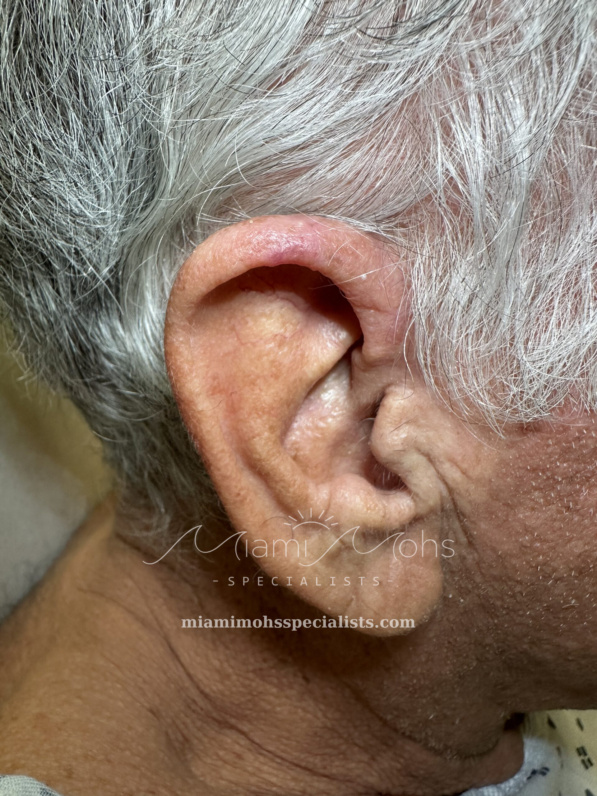
This gentleman presented with a nodular basal cell carcinoma on the helical rim of his right ear. During Mohs micrographic surgery, the visible tumor was removed and examined under the microscope in real time, revealing microscopic tumor roots extending beyond what was visible to the eye. Four layers were required to fully clear the cancer, resulting in a larger defect than initially expected, with full-thickness skin loss and exposure of the underlying cartilage.
The patient specifically requested a simple, low-maintenance repair. Dr. Freedman used a modified V-Y musculocutaneous flap to reconstruct the defect in a single stage. The patient was thrilled with the results.
Mohs Surgery Case #5
About this case
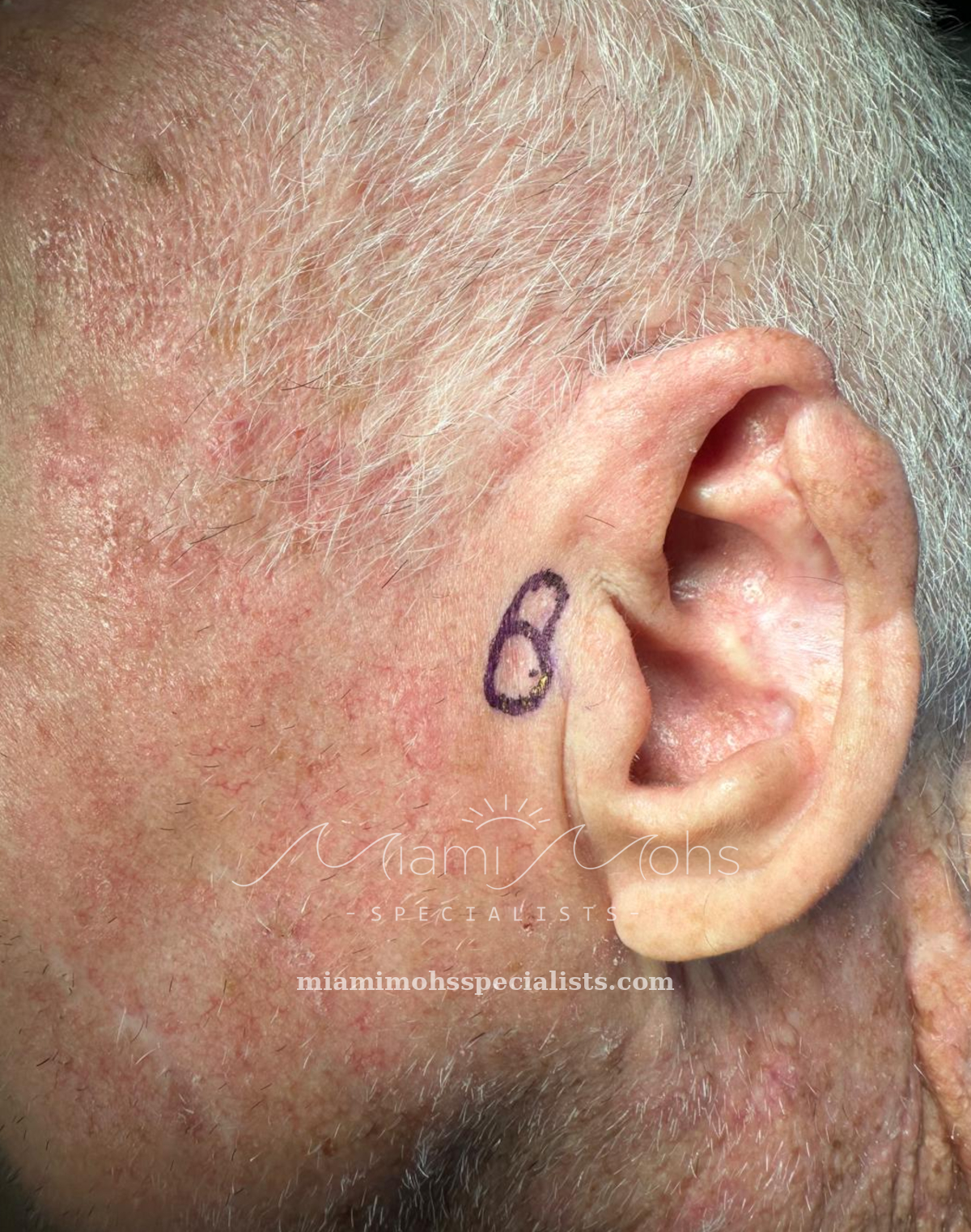
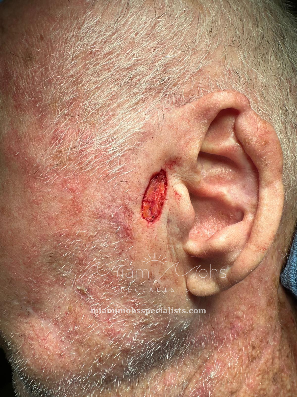
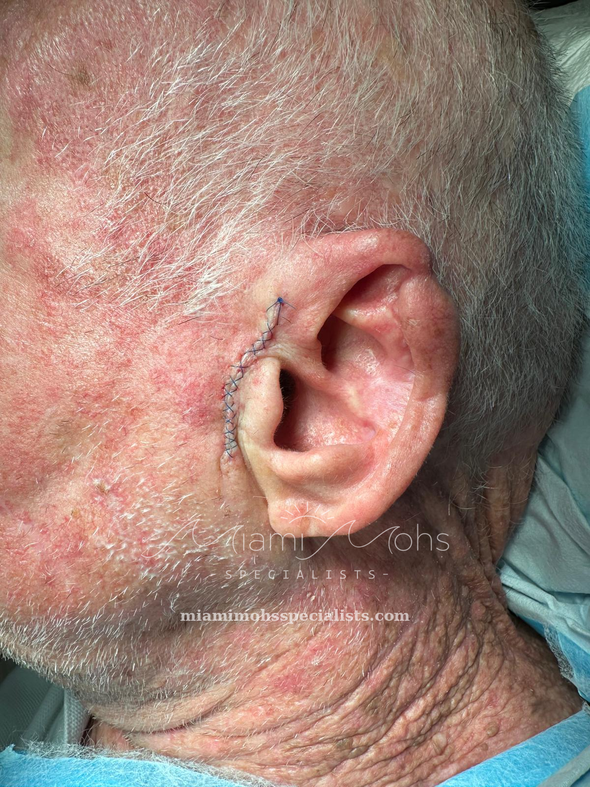
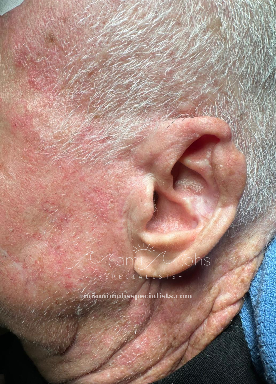
This case documents removal and reconstruction in the left preauricular area, immediately in front of the ear. The photographs show the area before Mohs surgery, the wound after Mohs surgery, and the completed reconstruction.
The final photograph was taken at the two-month follow-up and records the appearance during recovery. The sequence provides a view of this patient’s treatment and healing over time.